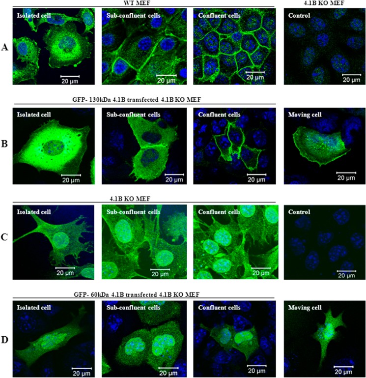
FIGURE 2.
Localization of 4.1B in MEF cells. A, localization of endogenous 130-kDa 4.1B. MEF cells were cultured on fibronectin-coated surface to reach different confluent conditions as indicated. The cells were then fixed and stained using anti-4.1B HP antibody. 4.1B KO MEF cells were used as the negative control. B, localization of exogenously transfected GFP-130-kDa 4.1B. GFP-130-kDa 4.1B was transiently expressed in 4.1B KO MEF cells. The cells were cultured as described, then fixed. The images were taken using confocal microscopy. C, localization of endogenous 60-kDa 4.1B. 4.1B KO MEF cells were cultured on fibronectin-coated surface to reach different confluent conditions as indicated. The cells were then fixed and stained using anti-4.1B exon 13 antibody. Cells stained with secondary antibody only were used as a negative control. D, localization of exogenously transfected GFP-60-kDa 4.1B. GFP-60-kDa-4.1B was transiently expressed in 4.1B KO MEF cells. The cells were cultured as described, then fixed. The images were taken using confocal microscopy. All pictures were taken with oil-immersed 63× objective. Scale bar, 20 μm.

