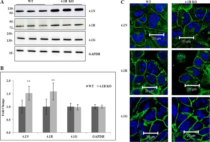
FIGURE 3.
Expression and localization of 4. 1N, 4.1R, and 4.1G in WT and 4.1B KO MEF cells. A, immunoblot analysis. Total lysates (40 μg protein) were probed with antibodies against 4.1N HP, 4.1R exon13, and 4.1G HP. GAPDH was used as the loading control. B, quantitative analysis of immunoblot. The results from three independent experiments were shown. **, p < 0.01. C, immunofluorescence staining. Cells were cultured on fibronectin-coated surface. One day after the cells reached confluence, the cells were fixed and stained using antibodies as indicated. All pictures were taken with oil-immersed 63× objective. Scale bar, 20 μm.

