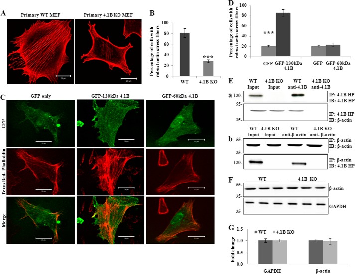
FIGURE 5.
130kDa-4.1B is required for actin stress fiber formation in MEF cells. A and B, endogenous actin filament staining. Cells were cultured on the fibronectin-coated surface and stained with Texas Red-phalloidin. The quantitative analysis from 50 cells of each cell type is shown in B. ***, p < 0.001. All pictures were taken with oil-immersed 63× objective. Scale bars, 20 μm. C and D, rescue of actin stress fiber formation by GFP-130-kDa 4.1B. 4.1B KO MEF cells were transfected with GFP, GFP-130-kDa 4.1B, or GFP-60-kDa 4.1B. Cells were stained with Texas Red-phalloidin. The quantitative analysis from 30 cells of each cell type is shown in D. ***, p < 0.001. All pictures were taken were taken with oil-immersed 63× objective. Scale bars, 20 μm. E, immunoprecipitation (IP). a, 130-kDa 4.1B was immunoprecipitated from MEF cells using anti-4.1B HP antibody. 130-kDa 4.1B or actin in the immunoprecipitate was detected using anti-4.1B HP antibody or anti-actin antibody. b, actin was immunoprecipitated from MEF cells using anti-actin antibody. Actin or 130-kDa 4.1B in the immunoprecipitate was detected using the indicated antibodies. IB, immunoblot. F and G, immunoblot analysis of actin. 20 μg of cell lysates were probed with anti-actin antibody. A GAPDH immunoblot is shown as a loading control (F). Quantitative analysis from three independent experiments is shown in G.

