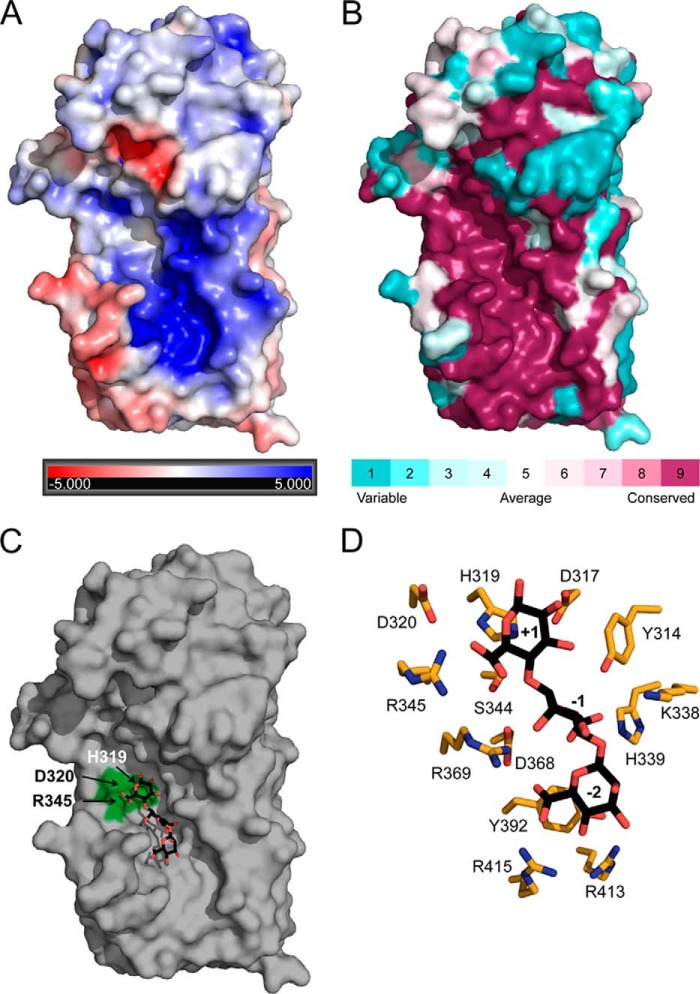
FIGURE 5.
Polymannuronate binding site. A, electrostatic potential surface representation of AlgG. The electrostatic properties of AlgG were determined with the APBS server. Positive surface potential is shown in blue, and negative surface potential is shown in red and contoured from −5 to +5 kT/e. B, surface representation of residue conservation of AlgG (dark magenta (high residue conservation) to dark cyan (variable residue conservation)). Residue conservation was produced with the ConSurf server using a T-Coffee alignment comprising AlgG sequences from Pseudomonas spp. and A. vinelandii. See “Experimental Procedures” for a list of sequences used. C, surface representation of AlgG with a modeled mannuronate trisaccharide in the putative poly(M) binding site. Residues His319, Asp320, and Arg345 are highlighted in green. D, stick representation of AlgG active and substrate binding site residues (orange) with modeled mannuronate trisaccharide (black).

