FIGURE 3.
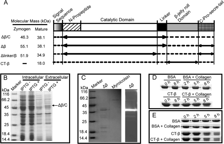
Expression and functional analysis of the truncated mutants of myroicolsin. A, schematic representation of the domain structures of myroicolsin and its mutants. The solid lines with arrows indicate the mutants' sequences, and the dotted lines indicate the deleted sequences. The predicted molecular masses from the amino acid sequence of each zymogen and mature protein are shown at the left. B, SDS-PAGE analysis of the overexpression of Δβ/C. The Δβ/C protein band is indicated by a black arrow. C, SDS-PAGE and gelatin zymography of Δβ. D and E, analysis of the collagen-binding ability of CT-β at 0 °C (D) and 25 °C (E). Collagen fibers (5 mg) were mixed with 20 μg of CT-β or BSA in 200 μl of buffer B and incubated at 0 or 25 °C. Samples were collected at 0, 2, 5, and 8 h and subjected to SDS-PAGE analysis. Collagen fibers incubated with BSA, CT-β incubated in buffer B, and BSA incubated in buffer B were used as the controls.

