
FIGURE 9.
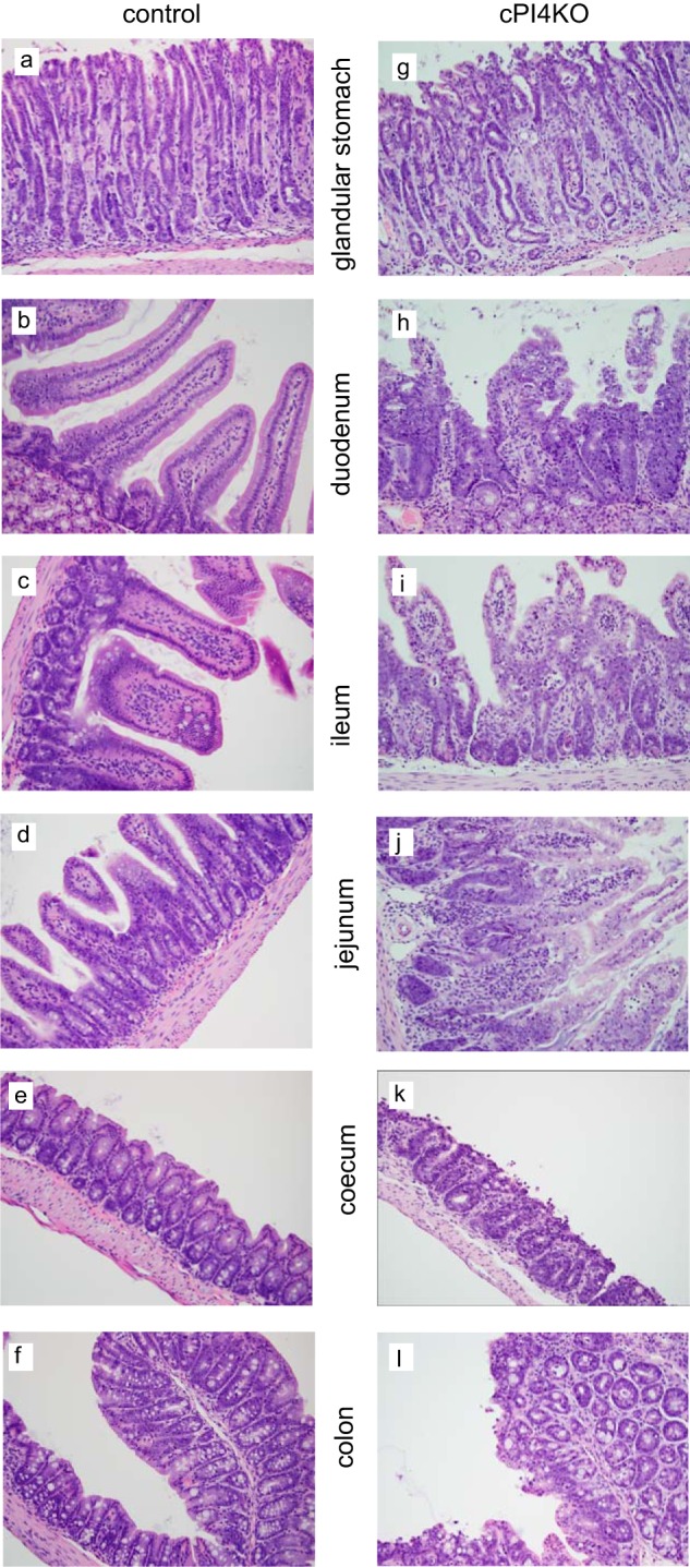
Histochemical analysis of selected segments of the GI track in wild-type and conditional pi4ka knock-out (cPI4KO) mice after tamoxifen treatment. Hematoxylin-eosin staining of the glandular stomach, duodenum, ileum jejunum, cecum, and colon. Panels a versus g, note the degeneration/necrosis of glandular pit, foveolar, and neck epithelial cells and lamina propria edema. Cellular debris is present within gastric pits in the cPI4KO animals. Panels b and c versus h and i, note the degeneration/necrosis of enterocytes on the villi and within intestinal crypts, crypt regeneration/hyperplasia (increased mitoses and cytoplasmic basophilia), and lamina propria edema in the cPI4KO animals both in the duodenum and ileum. Similar pathologies were observed in the jejunum, cecum, and colon (shown in panels d, e, and f versus panels j, k, and l).