FIGURE 2.

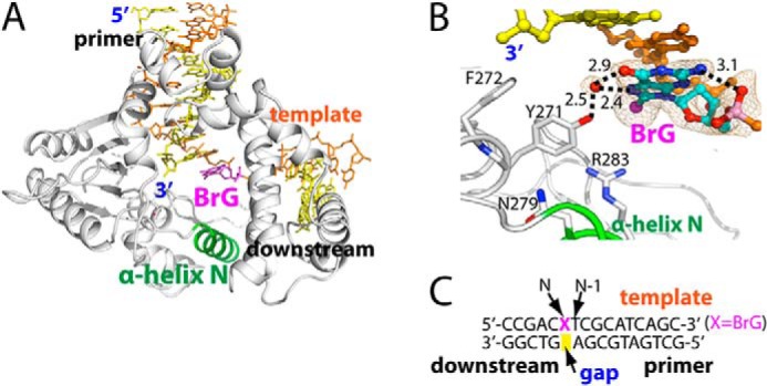
Structure of binary complex structure of polβ bound to DNA containing a single-nucleotide gap opposite templating BrG. A, overall structure of the binary gapped structure (PDB ID 4M2Y). Polβ is shown in white, and α-helix N containing the minor-groove recognition motif is shown in green. The template strand is shown in orange, and the primer and the downstream DNA are shown in yellow. BrG is shown in magenta. α-Helix N is in open conformation. B, active-site view of the gapped binary complex structure. An ordered water that bridges the OH of Tyr-271 and the Hoogsteen edge of BrG is depicted as a red sphere. Br is shown as a magenta sphere. Key H-bondings are indicated as dotted lines, where numbers in the panel indicate distances in Å. A 2Fo − Fc electron density map contoured at 1σ is shown around BrG and the ordered water. BrG is in syn conformation and forms an intramolecular H-bonding with its 5′-phosphate. C, DNA sequence used for crystallization of the gapped polβ complex. The positions of the gap, N, and N-1 are indicated.
