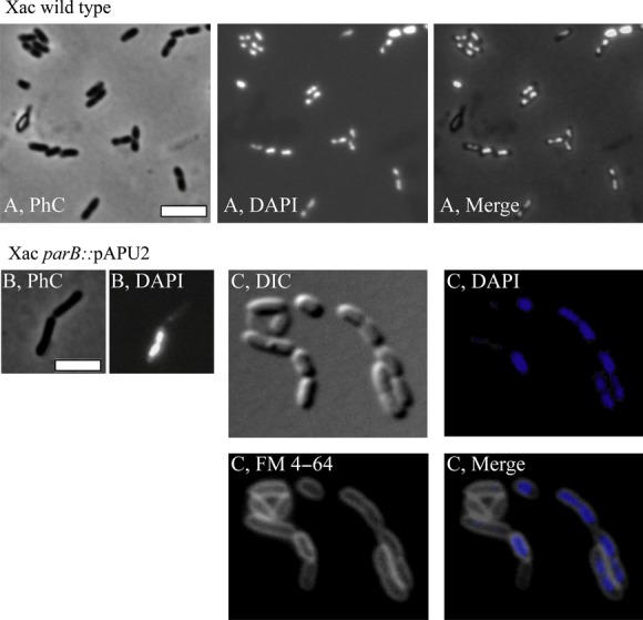
Figure 4.

Morphological analysis of Xac parB::pPAU2 mutant cells. The Xac parB::pPAU2 mutant, which expresses only truncated forms of ParB, was compared with Xac wild type using various combinations of phase contrast (PhC), differential interference contrast (DIC), and fluorescence microscopy; the membrane and nucleoid stains FM 4–64 and DAPI, respectively, were used as indicated. Wild-type and mutant cells were cultivated in NYG medium at 30°C, and inspected around the OD600 nm ˜0.3; cells were immobilized onto 1% agarose-covered slides. (A) Xac wild type. (B–C) Xac parB::pPAU2. Scale bar corresponds to 4 μm.
