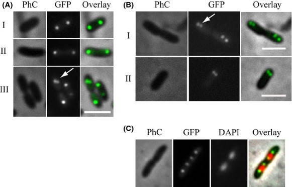
Figure 6.

Localization of ParB-GFP. The localization pattern of ParB-GFP was analyzed in different cell types of a Xac parB::pAPU3 expressing ParB-GFP from the native pparAB promoter. (A, B, C) represent cells photographed during different moments of the cell cycle; arrows mark recently divided origins of replication (see text). Cells were cultivated in NYG medium at 30°C until the OD600nm of ˜0.3, immobilized onto 1% agarose-covered slides, and visualized using fluorescence microscopy. Scale bar corresponds to 4 μm. PhC, phase contrast; DAPI, nucleoid stain.
