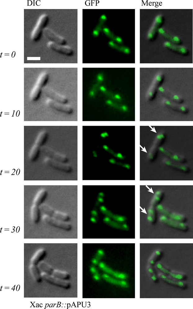
Figure 7.

Time-lapse microscopy of Xac parB::pAPU3. The localization of ParB-GFP in Xac parB::pAPU3 evidences an asymmetric mode of chromosome partitioning. Cells were cultivated in NYG medium at 30°C until the OD600nm of ˜0.3, and photographed at 0, 10, 20, 30, and 40 min as indicated. Arrows mark cells in which the replication origins have divided near the cellular poles (see text). DIC, differential interference contrast microscopy. Scale bar corresponds to 4 μm.
