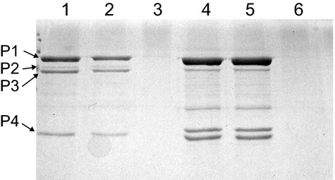
Figure 7.

SDS-PAGE chromatograph showing the four larger molecular-weight proteins (P1, P2, P3, and P4) of phage φ6. The lanes are as follows: (1) isolated φ6 virions; (2) MMT–φ6 virion pellet; (3) MMT-φ6 virion supernatant isolated from pellet preparation; (4) isolated NC; (5) MMT-NC pellet; and (6) MMT-NC supernatant isolated from NC pellet preparation.
