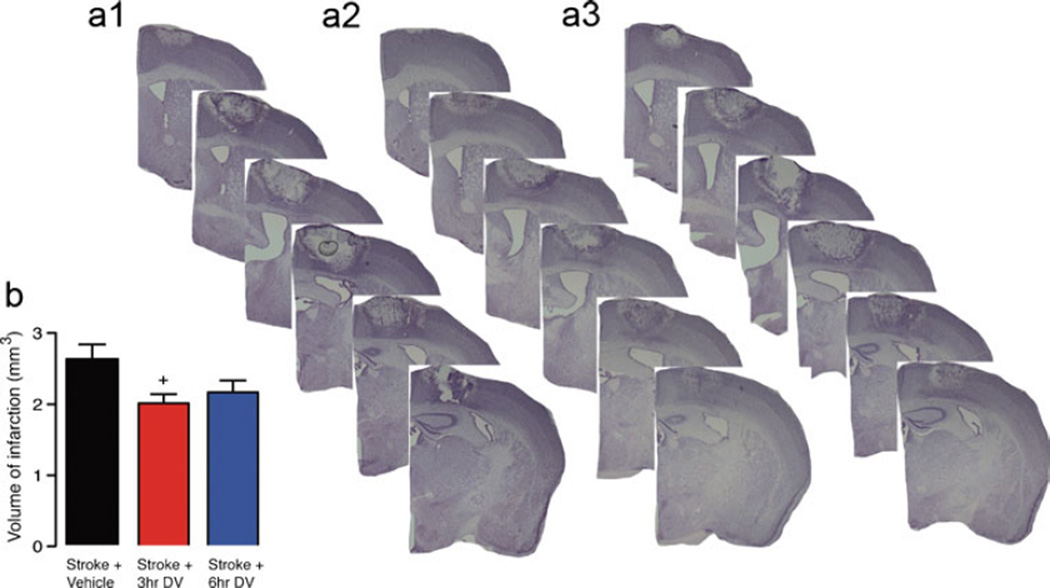
Fig. 2.
Histological assessment after stroke in aged. Representative cresyl violet-stained sections were generated 7 days post-stroke from vehicle-treated aged stroke control (a, 1) and stroke+DV treatment starting from 3 h (a, 2), and 6 h (a, 3) after stroke. Daily IP administration of DV (2 mg/kg) resulted in a significant decrease in infarct volume when given from 3 h post-stroke. Tissue was collected and processed 7 days post-stroke to quantify lesion size as shown in panel b. A n=5 per group were used. +=P<0.05 compared with stroke+vehicle-treated controls

