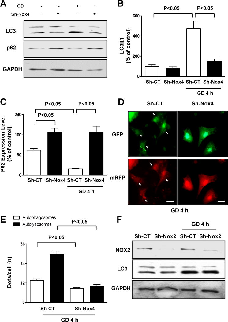
Figure 3. Nox4 is required for autophagy activation in CMs during GD.
A–C. CMs were transduced with Sh-CT or Sh-Nox4 for 96 hours, and then CMs were cultured with normal or glucose-free medium. LC3 and p62 protein levels were evaluated. Representative immunoblots (A) and densitometric quantification (B–C) are shown. N=4. Data are presented as a percentage of Sh-CT. D–E. CMs were transduced with Sh-CT or Sh-Nox4 for 96 hours together with Ad-mRFP-GFP-LC3 for the last 48 hours. CMs were cultured with glucose-free medium. Representative images of mRFP and GFP dots are shown (D), together with quantification of autophagosomes and autolysosomes (E). N=5. Bar=10 μm. F. CMs were transduced with Sh-CT or Sh-Nox2 for 96 hours, and then CMs were cultured with normal or glucose-free medium. LC3II and Nox2 levels were evaluated.

