Fig. 3.
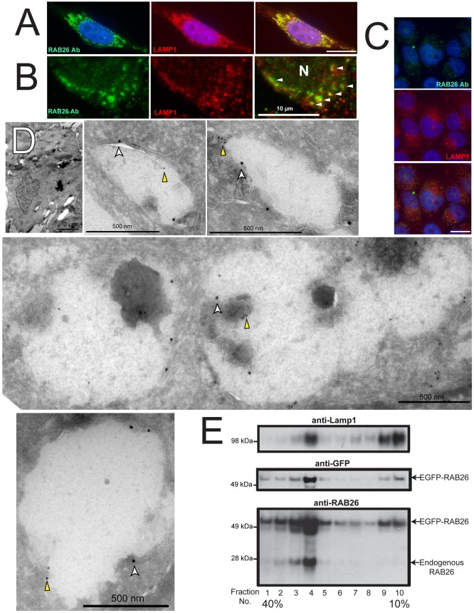
Endogenous RAB26 is also lysosome-associated. (A) Immunofluorescence imaging of endogenous RAB26 staining (green) in a rare HGC-27 cell with moderate RAB26 levels, co-stained for LAMP1 (red). (B) Confocal immunomicroscopy of endogenous RAB26 (green) and LAMP1 (red) (N, nucleus). Arrowheads indicate colocalization of RAB26 and LAMP1 vesicles. (C) Lower magnification epifluorescence images of HGC-27 cells with more representative, low-level endogenous RAB26 (green) and LAMP1 (red). (D) Immunoelectron micrographs of control GFP-transfected HGC-27 cells. A representative cell is shown at low magnification with several panels showing electron lucent structures labeled with anti-LAMP1 antibody (18-nm gold particles, white arrowheads) and sparsely labeled with anti-RAB26 antibody (12-nm gold particles, yellow arrowheads). (E) Density gradient cell fractionation experiment with numbered density fractions analyzed for lysosomes with anti-LAMP1 antibody, EGFP-tagged RAB26 by both anti-GFP and anti-RAB26 antibodies (middle and lower panel), and endogenous RAB26 by anti-RAB26 immunoblotting (lower panel). Scale bars: 20 µm unless indicated.

