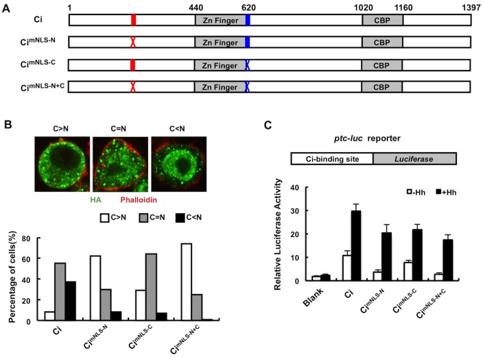
Fig. 2.
NLS-N acts in conjunction with NLS-C to regulate Ci nuclear localization and activity. (A) HA–Ci and its variants with NLS-N, NLS-C or both mutated. Red and blue bars represent wild-type NLS-N and NLS-C, respectively, whereas red and blue crosses represent mutated NLS-N and NLS-C, respectively. (B) S2 cells transfected with HA–Ci, HA–CimNLS-N, HA–CimNLS-C or HA–CimNLS-N+C were treated with 10 ng/ml LMB for 2 hours prior to immunostaining with an anti-HA antibody (green) and Phalloidin (red). Transfected cells show different patterns of nuclear and cytoplasmic distribution of tagged Ci or mutants. Quantification of nuclear and cytoplasmic localization of HA–Ci and its variants is shown below. (C) ptc-luc reporter structure and ptc-luc reporter assay in S2 cells expressing Ci or its variants in the absence or presence of Hh-conditioned medium. The y-axis represents normalized ptc-luc activity. Values are means ± s.d.

