Fig. 6.
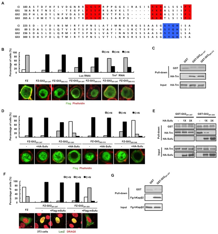
NLS-N is conserved in Gli proteins and regulated by Sufu. (A) Sequence alignment of the N-terminal conserved region of Ci/Gli (human Gli1, mouse Gli2 and human Gli3) with the PY-NLS motif highlighted in red and the Sufu binding core highlighted in blue. (B) Quantification of nuclear and cytoplasmic localization of FZ and its variants. Representative images of S2 cells transfected with FLAG–LacZ (FZ), FZ–Gli2201-247 and FZ–Gli3265-312 without or with control or Trnc dsRNA treatment and immunostained with anti-FLAG antibody (green) and Phalloidin (red) are shown below. (C) GST pull-down assay to show that HA–Trn is associated with GST–Gli2201-247 and GST–Gli3265-312. Input and bound HA–Trn proteins were visualized by western blot, whereas GST fusion proteins were detected by Coomassie Blue staining. (D) Quantification of nuclear and cytoplasmic localization of FZ and its variants. Representative images of S2 cells transfected with the indicated FZ–Gli chimera either alone or together with Sufu and immunostained with an anti-FLAG antibody (green) and Phalloidin (red) are shown below. Nuclear translocation of FZ–Gli2201-283 or FZ–Gli3265-348 but not FZ–Gli2201-247 and FZ–Gli3265-312 was blocked by Sufu coexpression. (E) GST pull-down assay to show that increasing amount of HA–Sufu blocked the binding of HA–Trn to GST–Gli2201-283 but not to GST–Gli3201-247. (F) Quantification of nuclear and cytoplasmic localization of FZ and its variants. Representative images of NIH3T3 cells transfected with the indicated FZ–Gli2 chimera either alone or together with mouse Sufu (mSufu) and immunostained with an anti-LacZ antibody (green) and DRAQ5 nuclear dye (pseudo red) are shown below. (G) GST pull-down assay to show that HA–hKapβ2 is associated with GST–Gli2201-247. Input and bound HA–hKapβ2 proteins were visualized by western blot whereas GST fusion proteins were detected by Coomassie Blue staining.

