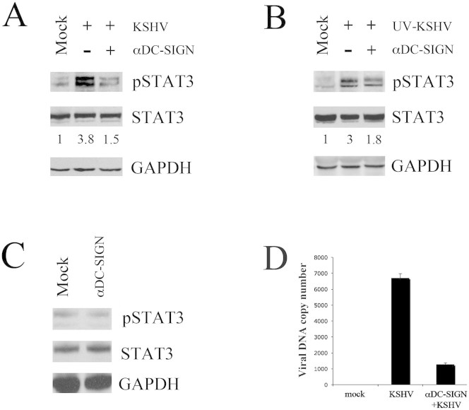
Figure 2. STAT3 activation by KSHV is partially dependent on viral engagement of DC-SIGN.
DCs were mock-treated or pre-incubated with anti-DC-SIGN (20 μg/ml) for 1 hr at 4°C, before exposure for 15 min at 37°C to (A) active KSHV or (B) UV-KSHV. STAT3 phosphorylation and its fold of induction, based on densitometric analysis of the ratio of phospho/total STAT3, are shown. (C) DCs were incubated with anti-DC-SIGN antibody alone for 15 min at 37°C and STAT3 phosphorylation was analyzed by western blotting. Total STAT3 and GAPDH were included as control. 1 × 10⁶/point overnight serum-starved DCs were used in all experiments. One representative experiment out of three is shown. (D) Real-time PCR showing KSHV DNA copy number/105 mock and KSHV-exposed DCs, pre-treated or not pre-treated with anti-DC-SIGN (20 μg/ml). Viral exposure was performed for 15 min at 37°C. Mean ± SD of three experiments is reported.

