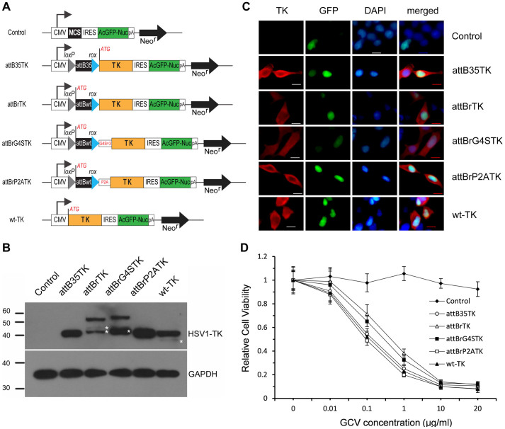
Figure 1. Suicide effect of different fusion TK proteins.
(A) Schematic of different fusion TK constructs. CMV: Cytomegalovirus immediate early promoter; loxP and rox: recognition targets of Cre and Dre recombinases; attB35 and attBwt: minimal fuctional size and full-length of wild-type attB site; G4S × 3: (Gly4Ser)3 flexible linker; P2A: self-cleaving 2A peptide derived from porcine teschovirus-1; ATG: initiation codon flanked by a Kozak consensus sequence; control: empty vector pIRES2-AcGFP1-Nuc. (B) Western blot analysis of HEK293 cells transfected with different TK constructs. The blot was probed with polyclonal antibodies to HSV-1 TK or GAPDH. The stars denote probable proteolytic breakdown products. Full-length blots are presented in Supplementary Figure S8. (C) Immunofluorescence staining of HEK293 cells at 48 h post-transfection. The nuclei were stained with DAPI (blue); TK proteins (red) were stained with an anti-TK antibody and visualized with Cy3-labeled secondary antibodies. Scale bars = 10 μm. (D) Relative viability of HEK293 cells transfected with different TK constructs. The transfected cells selected by FACS were exposed to different concentrations of the nucleoside analog GCV for 4 d. Cytotoxicity was assessed by the WST-1 assay. “Control” represented the cells transfected with the empty vector pIRES2-AcGFP1-Nuc.

