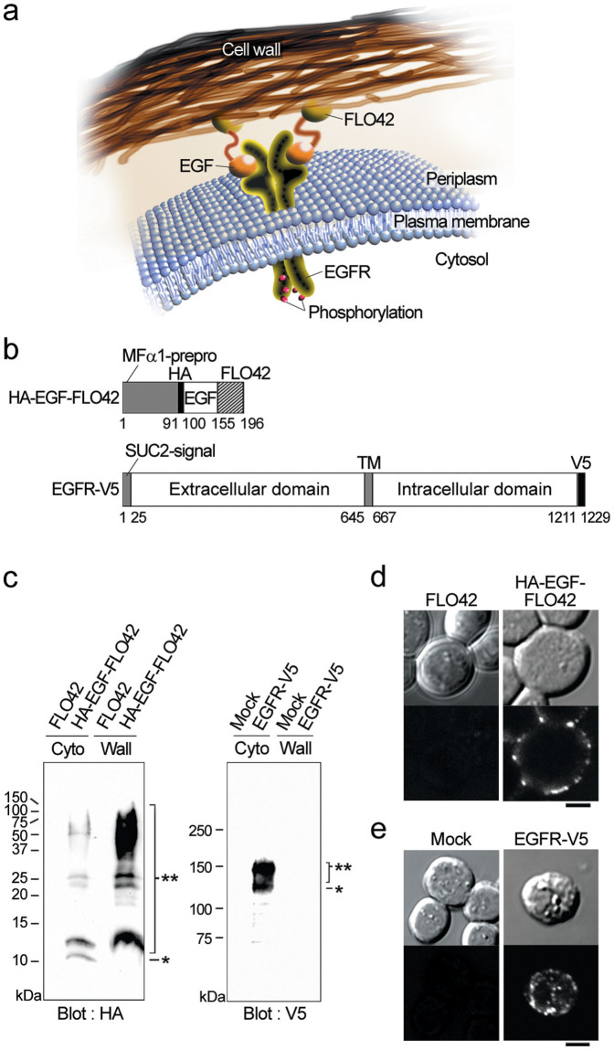
Figure 1. Expression and subcellular localization of the EGF-FLO42 and EGFR in yeast cells.
(a) Schematic drawing of the EGFR and FLO42-fused EGF in the periplasm of yeast cells. Upon binding EGF, the EGFR autophosphorylates. (b) A cell wall-anchored form of HA-tagged EGF (HA-EGF-FLO42) was expressed with the assistance of MFα1-prepro peptide. V5-tagged human EGFR (EGFR-V5) was expressed with the assistance of SUC2-signal peptide. TM, transmembrane domain. Numbers indicate amino acid residues at the borders of each domain. (c) Co-expression of HA-EGF-FLO42 and EGFR-V5 in yeast cells. Cytoplasmic (Cyto) and cell-wall (Wall) fractions were analyzed by western blotting using anti-HA (HA) and anti-V5 (V5) antibodies. Single and double asterisks indicate non-glycosylated and glycosylated forms, respectively. (d) Subcellular localization of HA-EGF-FLO42 in yeast cells. Intact cells were stained with anti-HA antibody. Bars = 2 μm. (e) Subcellular localization of EGFR-V5 in yeast cells. Spheroplasts were stained with anti-V5 antibody. Bars = 2 μm.

