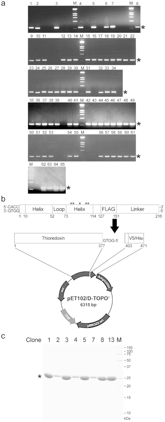
Figure 4. Single-cell PCR cloning and bacterial expression of HLH proteins.

(a) Single-cell PCR. PCR products obtained by single-cell PCR were subjected to agarose gel electrophoresis. Numbers indicate clones amplified successfully (65 clones/83 lanes). M, DNA marker. Asterisks indicate the position of target bands. (b) Directional TOPO cloning. The cDNA fragments amplified by single cell PCR (216 bp) were extracted from agarose gel, and then inserted into pET102 directional TOPO vector for the bacterial expression. Loop encodes hepta-glycine. Linker encodes N-terminal 20 amino acids of FLO42. Asterisks indicate the position of identical codon. (c) Purification of HLH proteins. HLH proteins expressed as an N-terminal thioredoxin-fused and C-terminal hexahistidine-tagged form in Escherichia coli BL21 (DE3) and purified with cobalt-chelating column. The purity was checked by CBB staining. Numbers indicate the clone number. M, protein marker. Asterisk indicates the position of target band.
