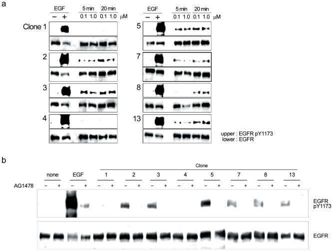
Figure 5. Induction of EGFR autophosphorylation in A431 cells by EGFR agonist candidates.
(a) After treatment with 0.1 and 1.0 μM bacterially expressed HLH peptides (8 species, clones 2 ~ 17) for 5 and 20 min, A431 cell lysates were subjected to western blotting with anti-EGFR and anti-EGFR pY1173 antibodies. As a positive control, 1.5 nM EGF was used. (b) After 30-min treatment with 200 nM tyrphostin AG1478, A431 cells were treated with 2.0 μM HLH peptides for 20 min, lysed and subjected to western blotting with anti-EGFR and anti-EGFR pY-1173 antibodies. As a positive control, 1.5 nM EGF was used.

