
Fig. 1.
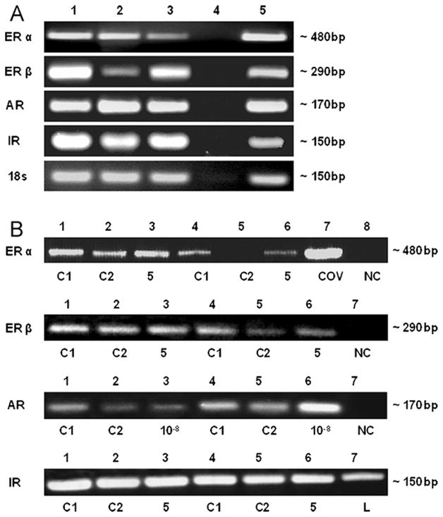
Detection of estrogen receptors (ER) α and β, androgen receptor (AR) and insulin receptor (IR) transcripts in chondrocyte cell lines C-28/I2 and T/C-28a2 and in human primary articular chondrocytes cultured without hormones (A) and with hormones (B). (A) ER alpha (483 bp) and beta (295 bp), AR (168 bp) and IR (152 bp) mRNAs were detected in human primary articular cartilage cells (lane 1), C-28/I2 and T/C-28a2 chondrocytes (lanes 2, 3) and in uterine cells used as positive control (lane 5). DEPC-water served as negative control (lane 4). As further control, RT-PCR analysis of the household gene 18s was performed. PCR bands were shown by means of agarose gel electrophoresis. (B) Untreated controls were C1 (serum-free medium) and C2 (serum-free medium with solvent, PBS/NaOH). DEPC-water as negative control (NC) is indicated. ERβ mRNA (483 bp) levels were analyzed by RT-PCR in C-28/I2 cells incubated without (lanes 1, 2, 4, 5) or with 5 μg/ml insulin (lanes 5, 6), compared to total RNA extracted from human granulosa cells (COV434) as positive control (lane 7). ERβ mRNA (295 bp) levels were analyzed in C-28/I2 cells incubated without (lanes 1, 2, 4, 5) or with 5 μg/ml insulin (lanes 3, 6). AR mRNA (168 bp) was analyzed in T/C-28a2 cells incubated without (lanes 1, 2) or with 10−8 M dihydrotestosterone (lane 3) and in C28/I2 cells incubated without (lanes 4, 5) or with 10−8 M dihydrotestosterone (lane 6). IR mRNA (152 bp) was analyzed in C-28/I2 cells incubated without (lanes 1, 2, 4, 5) or with 5 μg/ml insulin (lanes 3, 6), compared to total RNA extract from liver (L). PCR bands were shown by means of agarose gel electrophoresis.