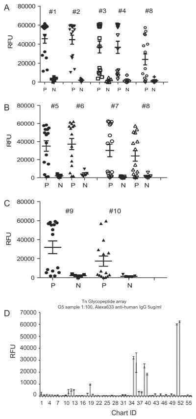
Fig. 1.
Reactivity to Cryptosporidium parvum 17 kDa synthetic peptides with different Tn glycosylation. (A) Differences in serum responses to the glycosylated Cp17 peptides #1–4 compared with non-glycosylated peptide #8 (P < 0.04, 0.04, 0.04 and 0.05, respectively, compared with #8). (B) Responses to the glycosylated Cp17 peptides #5–7 compared with non-glycosylated peptide #8 (P < 0.07, 0.4 and 0.075, respectively). (C) Responses to the glycosylated Cp17 peptide #9 compared with non-glycosylated peptide #10 (P < 0.06). Sera from cryptosporidiosis patients who were antibody positive (P) (n = 15), as well as from antibody-negative healthy blood donors (N) (n = 5) were diluted 1:100 and analysed for the presence of anti-IgG antibodies using the printed array of synthesised Cp17 peptides (Table 1). (D) Representative example of Tn glycopeptide array data for one patient serum sample. RFU, relative fluorescent units. See Supplementary Table S1 for complete data set. Chart ID numbers 12, 13, 19 and 35–40 where binding is observed all contain varying numbers and presentations of the Tn antigen.

