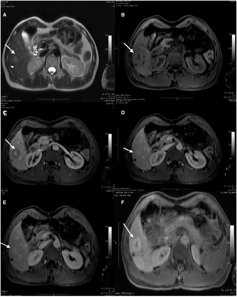
Figure 2.
Contrast enhanced Magnetic Resonance Imaging (CEMRI), showing two focal liver lesions consistent with peliosis. T2-weighted (A), T1-weighted volume interpolated body examination (VIBE) (B), T1-weighted arterial phase (C), T1-weighted portal venous phase (D), T1-weighted late venous phase (E) and T1-weighted hepatobiliary phase (F) showing a focal liver lesion (arrows) slightly hyperintense on T2-weighted images with hypointense signal on T1-weighted images and centrifugal contrast enhancement and peripheral contrast media uptake on hepatobiliary phase.

