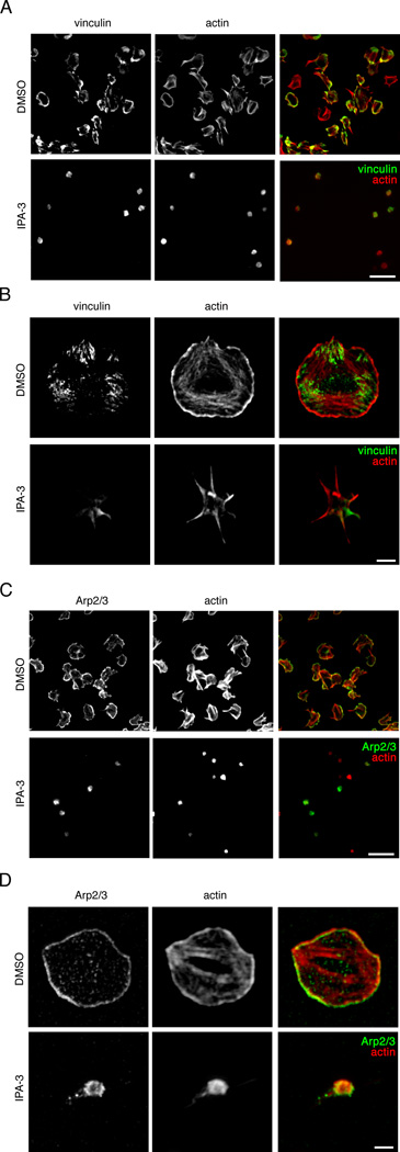
Figure 5. PAK activity regulates actin-rich adhesion and lamellipodia formation in platelets spread on collagen.
Replicate wells of collagen-coated coverglass were seeded with human platelets (2 × 10⁷/ml) for 45 min after pretreatment with vehicle (DMSO), IPA-3 (10 µM), PIR 3.5 (10 µM) or PF-3758309 (10 µM). Platelets were fixed, stained for (A,B) vinculin and actin or (C,D) Arp2/3 and actin and analyzed by fluorescence microscopy. Images representative of five experiments are shown. (A,C) Scale bar = 10 µm. Super resolution microscopy analysis of (B) platelet focal adhesion formation and (D) lamellipodia formation of vehicle (DMSO) and IPA-3 (10 µM) treated platelets on a surface of collagen, visualized by SR-SIM of actin (red) and vinculin or Arp2/3 (green) staining. Scale bar = 2 µm.

