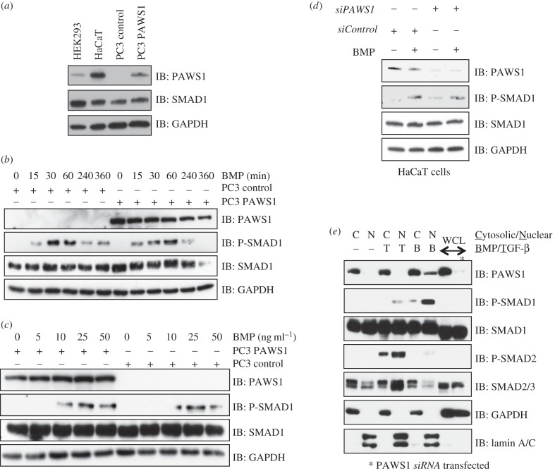
Figure 3.
Effect of PAWS1 on BMP-induced SMAD1 phosphorylation. (a) Extracts (20 μg protein) from either HEK293, HaCaT, PC3-control (PC3 cells stably integrated with a control vector), or PC3-PAWS1 cells (PC3 cells stably integrated with a vector encoding wild-type PAWS1) were resolved by SDS–PAGE and analysed by immunoblotting using the indicated antibodies. (b) PC3-control and PC3-PAWS1 cells were treated with BMP-2 (25 ng ml−1) for the indicated time (min) prior to lysis. Extracts (20 μg protein) were resolved by SDS–PAGE and analysed by immunoblotting using the indicated antibodies. (c) PC3-control and PC3-PAWS1 cells were treated with the indicated concentrations of BMP-2 for 1 h prior to lysis. Extracts (20 μg protein) were resolved by SDS–PAGE and analysed by immunoblotting using the indicated antibodies. (d) PAWS1-depleted HaCaT cells (siPAWS1) and HaCaT cells expressing FOXO4 siRNA (siControl) were treated either with or without BMP-2 (25 ng ml−1) for 1 h prior to lysis. Extracts (20 µg protein) were resolved by SDS–PAGE and immunoblotted with the indicated antibodies. (e) Extracts from HaCaT cells treated with either BMP-2 (25 ng ml−1) or TGF-β (50 pM) for 1 h, or left untreated were separated into cytosolic and nuclear fractions. Fractions and whole cell lysates (WCLs) were resolved by SDS–PAGE and analysed by immunoblotting using the indicated antibodies. GAPDH and lamin A/C were used as cytosolic and nuclear controls, respectively.

