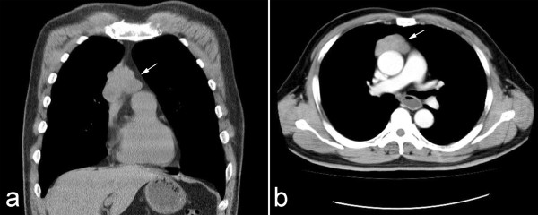
Figure 1.

Preoperative chest radiology of lesion. (a) Chest CT image showed a well-defined mass in right anterior mediastinum (white arrow). The tumor was observed to adhere to the wall of great vessel. (b) The contrast enhanced CT scan showed the solid mass with mild heterogeneous enhancement adhered, but not invaded the wall of great vessel.
