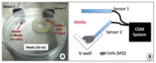
Figure 3. Sensor-cell culture system used to evaluate the impact of cell populations on glucose sensor function in vitro.

The sensor-cell culture system presented in Figure 3a and 3b were developed to mimic the cell sensor interactions that occur at the sensor-tissue interface at glucose sensor implantation sites.
Figure 3a is a photograph of our dual sensor cell culture system for evaluation of the impact of cell populations on glucose sensor function and CGM in vitro. The Cell culture system is composed of a standard tissue culture petri dish in which a nylon disk with a V-well drilled into one edge of the nylon disk. Additionally, 2 sensors are inserted into the petri dish thru predrilled holes, and sealed with aquarium silicone. For this system 50 ml of standard cell culture media is added to the petri dish, and candidate cells are pipetted into the V-well containing glucose sensor 2 (thin red arrow).
Figure 3b is a diagram of the V-well area in the nylon disk used in the dual sensor-cell culture system presented in Figure 3a. Also included is the location of the dual sensor system and the link to the CGM electronics.
