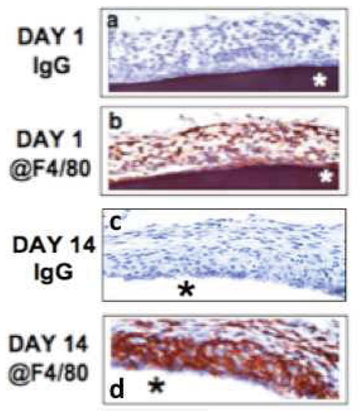
Figure 4. Characterization of macrophage distribution at glucose sensor-tissue interface at sites of glucose sensor implantation using anti-F4//80 antibodies.

Figures 4a–d are photomicrographs of IHC analysis of sensor-tissue interface at sites of glucose sensor implantation in our murine model of CGM. The goal of these studies was to characterized the presence and distribution of macrophages at sites of sensor implantation in the mouse using @F4/80 IHC. Using @F4/80 antibodies and standard IHC technology, scattered MQs were detected as early as 1-day post senor implantation (Figure 4b). Accumulation of MQ at the sensor-tissue interface continued to increase, ultimately forming a large MQ “band/barrier” which creates “Cell Based Metabolic Barrier” of macrophages surrounding the sensor at day 14-post implantation and beyond (Figure 4b). As a control normal Ig was tested at all time points and was negative for all time-points (Figure 4a and 4c).
