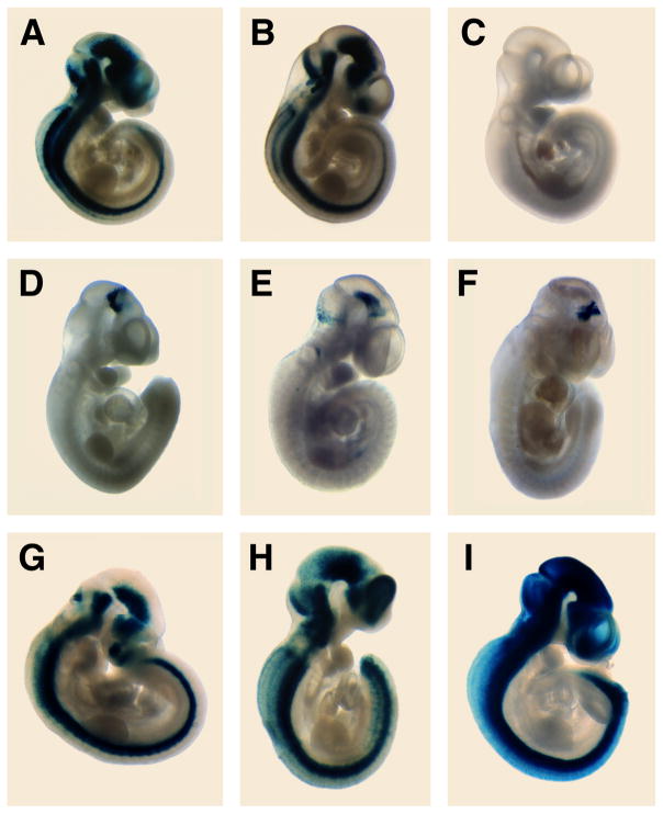
Figure 2. Whole mount β–galactosidase activity in transient transgenic embryos.
Embryos were isolated at 10.5 dpc and assayed for β–galactosidase activity. Reprentative embryos are depicted for A: construct 1; B: construct 4; C: construct 5; D: construct 8; E: construct 10; F: construct 11; G: construct 15; H: construct 19; I: construct 20. Consistently, three staining patterns emerged: the entire developing CNS was positive (A, B, G–I), or ventral midbrain only (D–F), or the neuroepithelium was negative (C).

