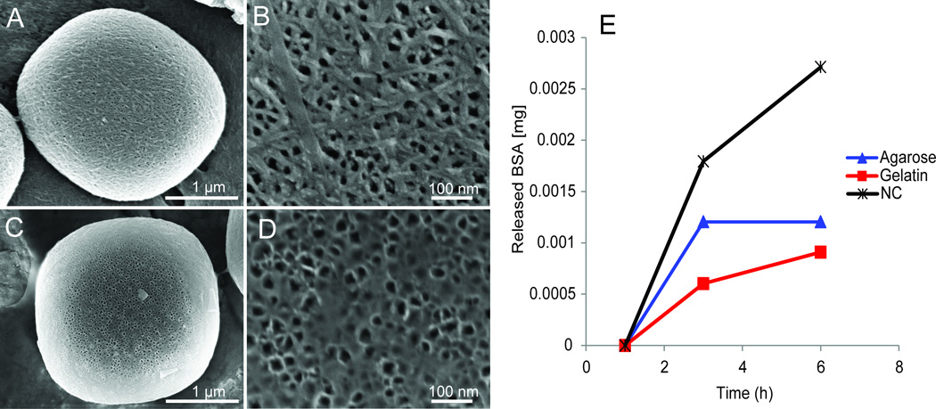
Figure 5. Coating and delayed release of biomolecules from MSN.
A–D. SEM micrographs demonstrating the coating of MSN with agarose (A,B) and gelatin (C,D) at the single particle scale (A,C) and pore scale (B,D). E. Release profiles of BSA-FITC from uncoated (black), agarose (blue), and gelatin (red) coated MSN. The coating of MSN yielded a delay in the release of BSA enabling kinetics not possible without a coating.

