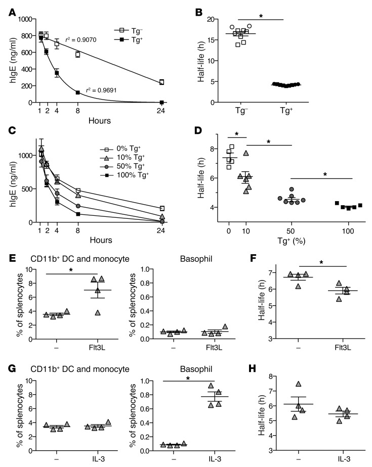
Figure 7. Human FcεRI expressed by DCs or monocytes significantly contributes to serum IgE clearance in FCER1A-Tg mice.
(A) Serum IgE clearance in Tg+ and Tg– littermates. 2.5 μg hIgE was i.v. injected into mice, blood was collected following injection, and hIgE concentration was determined by ELISA. Data are representative of 3 independent experiments using 3 mice per strain. For each mean, a line of best fit with indicated r2 values was calculated. (B) Serum half-life of hIgE in Tg+ and Tg– mice. IgE half-life was calculated as the time for peak serum IgE to halve from lines of best fit calculated as in A. Symbols represent individual mice injected with 2.5 μg (squares) and 5 μg (circles). *P < 1.5 × 10⁻⁶. (C and D) hFcεRI-expressing hematopoietic cells are responsible for serum IgE clearance. Mixed bone marrow chimeras were made on Tg– recipient hosts, and serum hIgE clearance was examined as described for A and B. *P < 0.05. (E and G) Flt3L increases the frequency of DCs/monocytes, while IL-3 increases basophils. 10:90 (Tg+/Tg–) mixed chimeric mice were injected with Flt3L-expressing B16 cells (E) or with IL-3/anti–IL-3 antibody complexes (G). The percentage of [CD11b+ DCs + monocytes] or basophils in spleen was determined by flow cytometry. *P < 0.05. (F and H) Serum hIgE clearance is accelerated by an increase in DCs/monocytes but not by an increase in basophils. *P < 0.05. Data in C–H are representative of 2 independent experiments. The second experiment of E–H is shown in Supplemental Figure 11.

