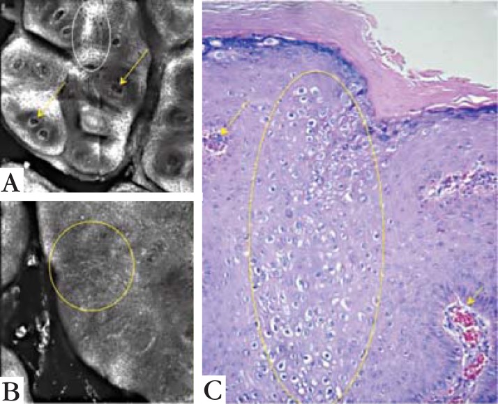
FIGURE 5.
Comparing confocal microscopy findings with the histopathologic image. (A) is a 1 x 1 mm view with vessels pointed out by the arrows. The same vessels are pointed on histologic image C. (B) is a 0.5 x 0.5 view showing the architectural disruption of the spinous layer without the usual honeycomb pattern. The disruption is seen on histologic image C as koilocytosis (yellow circles). On A it is also possible to see areas of the spinous layer with a honeycomb pattern (white circle)

