
Figure 2.
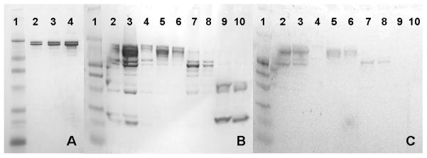
(A,B) Non-heated, non-reducing, SDS-PAGE gel stained for protein of the mAb in the 2.2 mM sodium phosphate, pH 6 formulation. (C) Same conditions as panels A and B except stained for glycoprotein (molecular weight marker is prestained for protein). The molecular weight marker in all three panels is in lane 1 and has bands at 210 kDa, 110 kDa, 80 kDa, 47 kDa, 32 kDa, 25 kDa and 16.5 kDa. Panel A consists of the control mAb (no incubation) in lanes 2–4 at loadings of 1, 2 and 5 μg protein. In panels B and C lanes 2–4 are the mAb incubation products after 50 days at 40°C as injected for SE-HPLC at three loadings. From left to right the loadings are approximately 5, 10 and 1 μg total protein respectively. Lanes 5 and 6 are the enriched monomer fraction collected during SE-HPLC (peak eluting at ~8.6 mL in Figure 1B) at loadings of 2 and 1 μg total protein respectively. Lanes 7 and 8 are the enriched large fragment fraction (fragment 1, the peak eluting at ~9.3 mL in Figure 1B) as collected from SE-HPLC at loadings of 2 and 1 μg total protein respectively. Lanes 9 and 10 are the enriched smaller fragment species that elute near 10.8 mL (fragment 3) as collected from SE-HPLC at loadings of 2 and 1 μg total protein respectively.