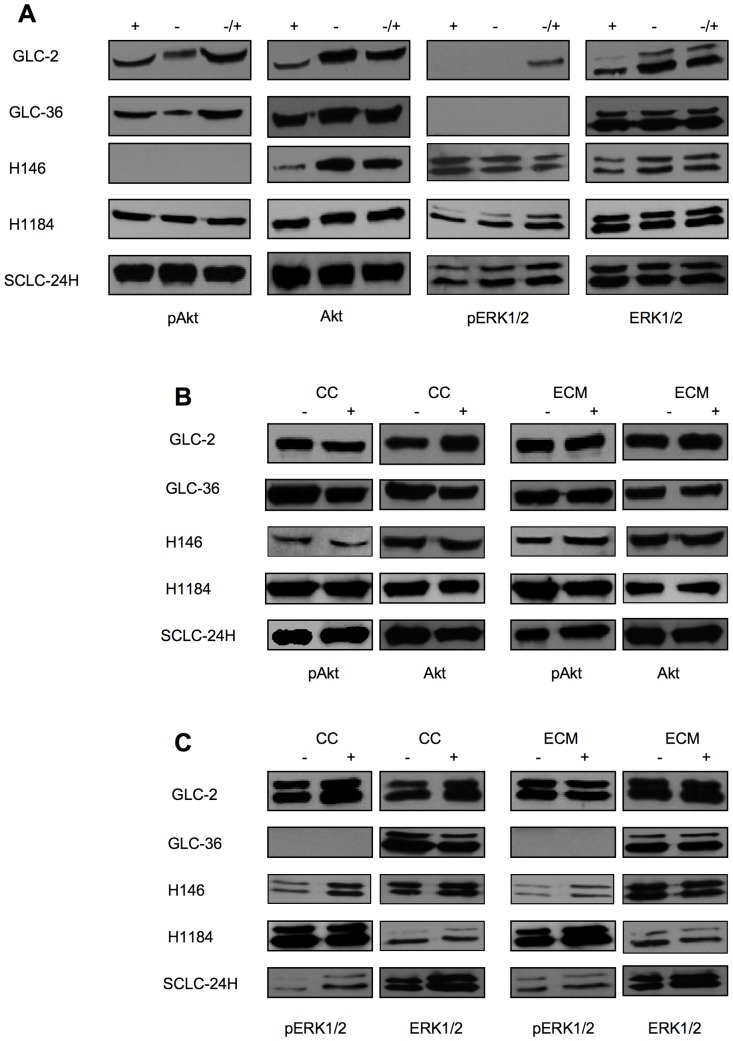
Figure 6. Western blot analysis of Akt and ERK1/2 activation in SCLC cells.
(A) SCLC cells were cultured for 24 h in the presence (+) or in the absence of serum (−) or in the presence of serum only during the last 10 min of the 24 h culture period (−/+). (B, C) SCLC cells were cultured for 24 h in the absence (−) or presence (+) of 50 nM SSP either on cell culture plastic (CC) or on ECM constituents, whereby FIB was used for all cell lines with the exception of SCLC-24H cells, that were cultured on a LAM substrate. Cell lysates were analyzed for the presence of total or phosphorylated (p) Akt (B) or ERK1/2 (C) proteins. Equal amounts of protein (20 µg) were loaded per lane.

