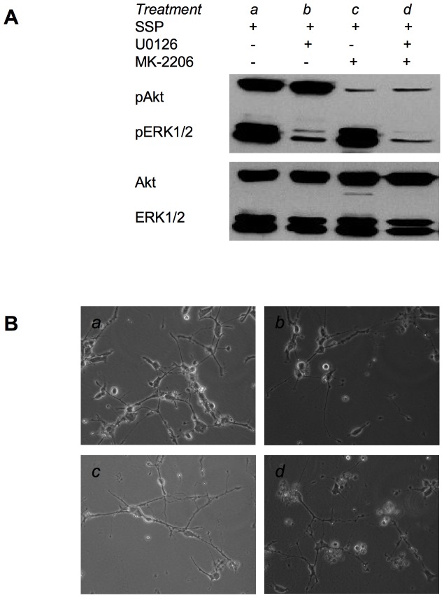
Figure 7. Pharmacological inhibition of Akt and ERK1/2 activation in SCLC-24H cells.
(A) Western blot analysis of cell lysates from SCLC-24H cells that had been cultured for 48 h on a LAM substrate in the presence (+) or absence (−) of 10 µM U0126 or MK-2206 and in the presence (+) of 20 nM SSP for the final 24 h of the culture period. Cell lysates were analyzed for the presence of total or phosphorylated (p) Akt or ERK1/2 proteins. (B) Micrographs of SCLC-24H cells that had been cultured under various conditions as described in (A) and small letters refer to the respective treatments. Equal amounts of protein (20 µg) were loaded per lane.

