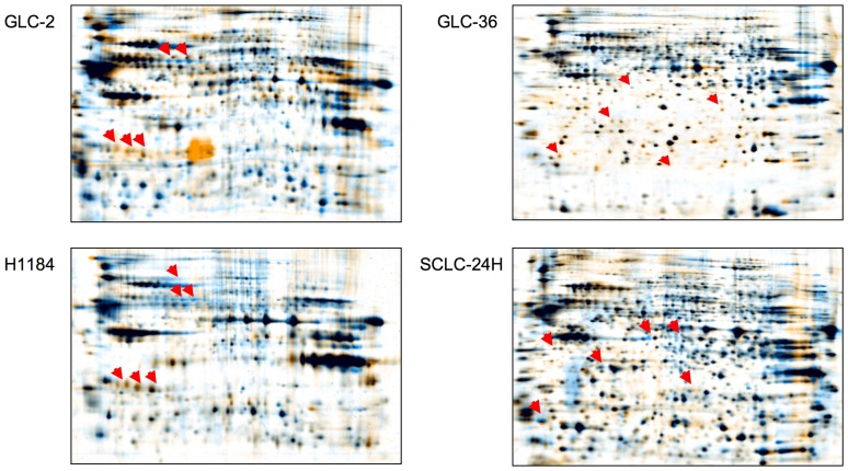
Figure 8. Comparative gel analysis of SCLC cell lysates.
Cells were cultivated for 30(blue) or absence (orange) of 20 nM SSP on a FIB (GLC-2, GLC-36, H1184 cells) or a LAM (SCLC-24H cells) substrate. Arrowheads indicate proteins that appear to be differentially expressed in SSP-treated versus untreated cells.

