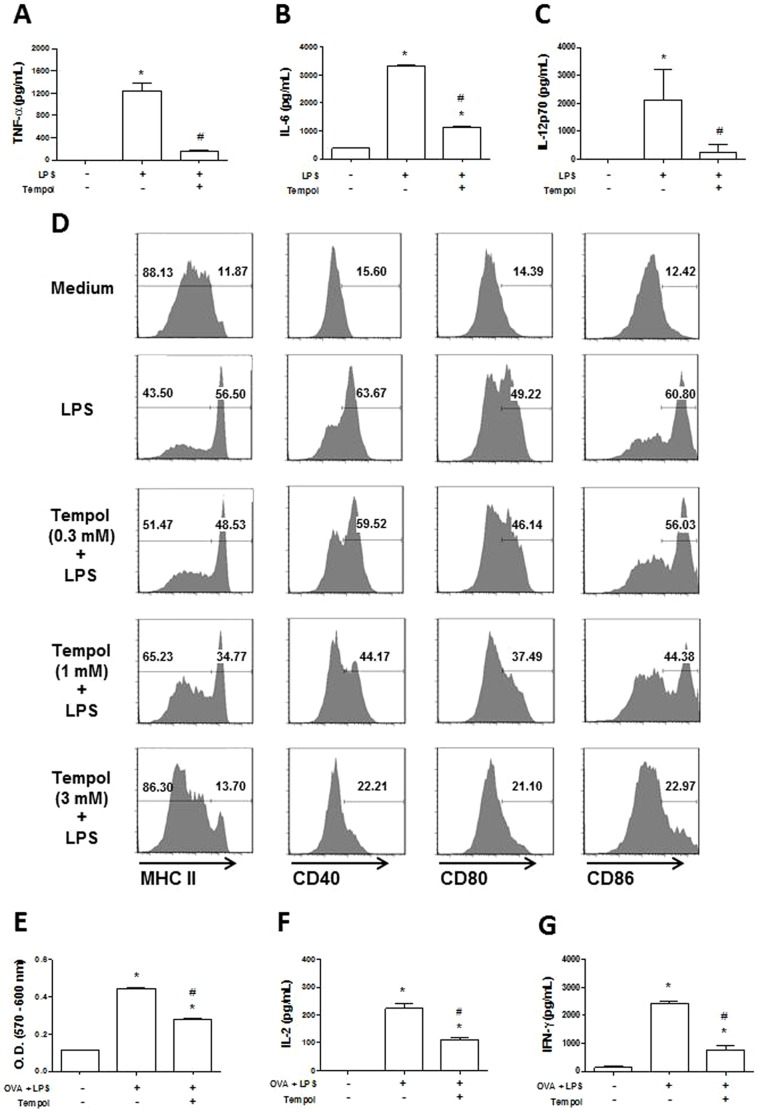
Figure 3. Tempol inhibits dendritic cell (DC) functions.
Bone marrow-derived DCs (106 cells/mL) were preincubated overnight with medium or Tempol at indicated concentrations and stimulated with lipopolysaccharide (LPS) (200 ng/mL) for 24 hours. Detection of A, TNF-α was evaluated in cell-free culture supernatants after six hours, and the levels of B, IL-6 and C, IL-12p70 were evaluated after 24 hours. D, Cells were stained with fluorochrome-labeled monoclonal antibodies to CD11c, CD40, CD80, CD86, and MHC class II and analyzed by flow cytometry. E, DCs incubated with medium or Tempol were pulsed with OVA (100 µg/mL) plus LPS (200 ng/mL) for four hours. After repeated washings, DCs were co-incubated with purified CD4+ cells from DO11.10 mice (DC:CD4+ ratio = 1:4) for 72 hours, and proliferation was measured as described in Materials and Methods. F, IL-2 and G, IFN-γ were evaluated in the culture supernatants from the proliferation assay. *, P≤0.05 versus control group (–/–); #, P≤0.05 versus LPS group (+/−) (analysis of variance).

