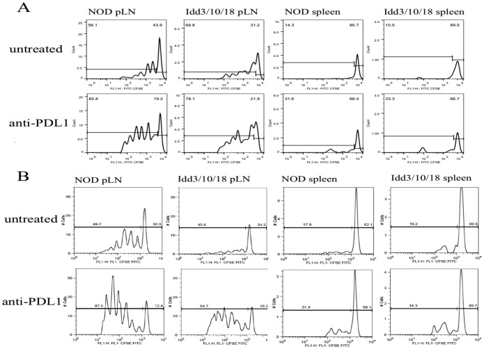
Figure 4. Proliferation of adoptively transferred BDC2.5 Tg CD4 T and NY8.3 tg CD8+ T cells in pancreatic LN and spleen of anti PDL1 treated NOD and Idd3/10/18 mice.
A) Representative CFSE dilution plot for each group is shown. Cells were gated on CD4+ Vbeta 4+. B) A representative CFSE dilution plot of transferred NY8.3tg T cells for each group is shown. Cells were gated on CD8+ Vbeta 8+.

