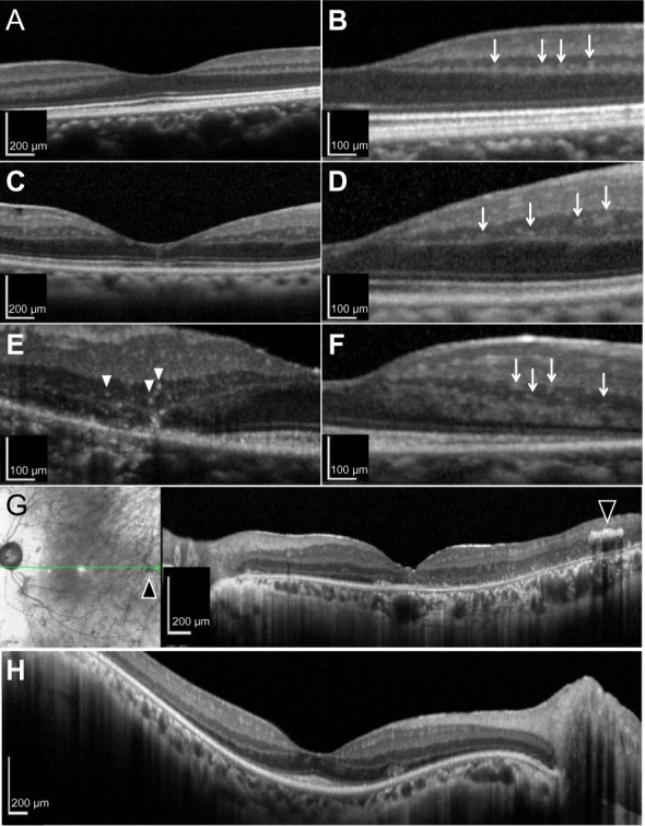
Figure 2.

HFs in the INL of normal and RP retinas. (A and B) HFs were located on the edge of the INL adjacent to the OPL in the normal retina (arrows). (C and D) HFs were located in the middle of the INL of the RP retina (arrows). HFs in the INL were of various sizes and signal intensities, ranging from relatively small and hyperreflective (E, arrowheads) to large and hyporeflective (F, arrows). (G) A high-intensity object was observed in the INL by OCT in a location homologous to bone spicule-like pigmentation (arrowheads). (H) In some cases with an IS/OS under the fovea, HFs were observed only in the ONL and not in the INL.
Abbreviations: HFs, hyperreflective foci; INL, inner nuclear layer; OPL, outer plexiform layer; IS/OS, inner segment/outer segment junction of photoreceptors; ONL, outer nuclear layer; RP, retinitis pigmentosa; OCT, optical coherence tomography.
