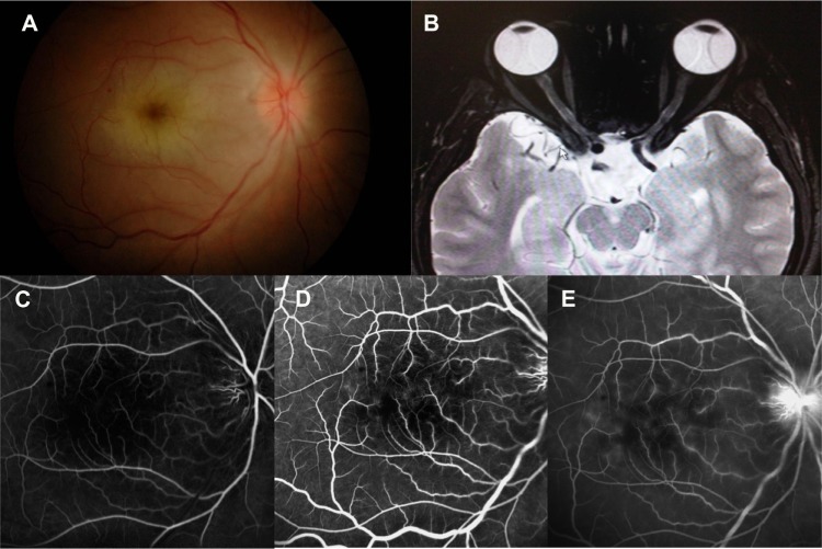
Figure 1.
(A) Fundus photograph at presentation showing optic disc edema, whitening and edema of the posterior pole, and a cherry red spot. (B) Increased signal in T2-weighted magnetic resonance imaging and enhancing with contrast in right optic nerve. (C–E) Fundus fluorescein angiogram showing capillary dropout area, minimal vascular and disc leakage.

