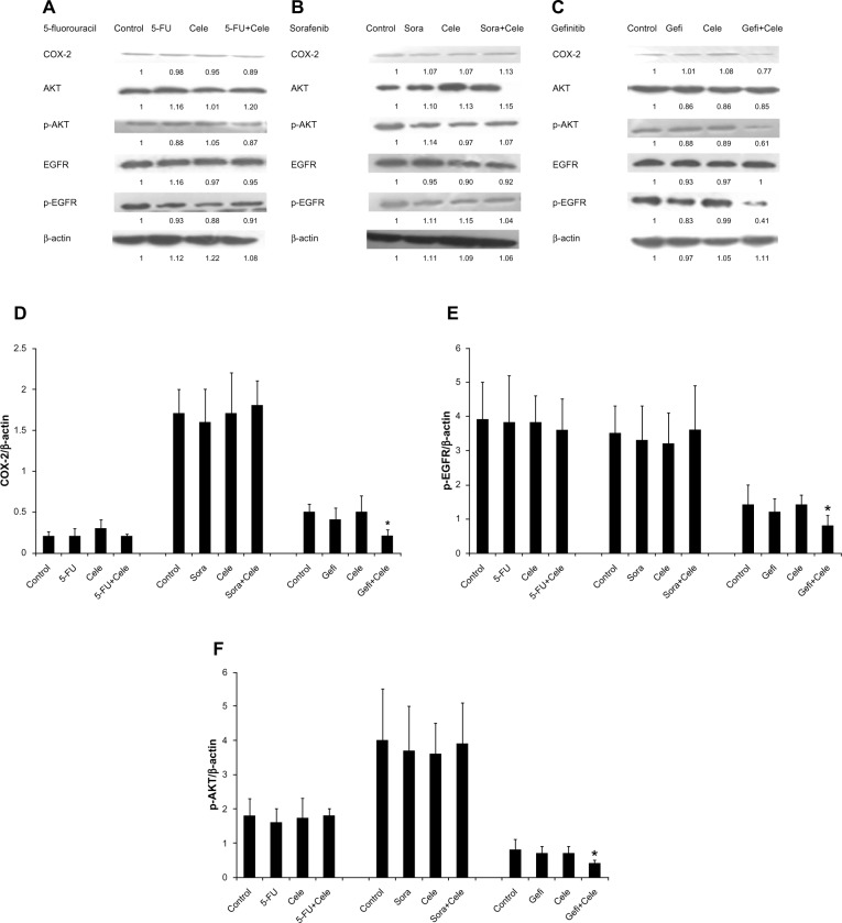
Figure 6.
(A–F) Protein expression of cyclooxygenase (COX)-2, phosphorylated epidermal growth-factor receptor (p-EGFR), and phosphorylated (p)-AKT in HepG2 multicellular spheroids determined by immunoblot analysis. After concurrent exposure of 48 hours with 5-fluorouracil (5-FU; 8.1×10⁻³ g/L), sorafenib (Sora; 4.4 μmol/L), and gefitinib (Gefi; 5 μM) with or without celecoxib (Cele; 21.8 μmol/L), the samples were collected. Samples incubated with only celecoxib were obtained 48 hours after celecoxib administration. (A–C) Immunoblots represent observations from one single experiment repeated three times. The integrated optical densities of (D) COX-2, (E) p-EGFR, and (F) p-AKT proteins were analyzed after normalization with β-actin (43 kDa) in each lane. (D–F) Means ± standard error of the mean of three separate experiments.
Note: *Statistical difference when compared to the control group (P<0.05).

