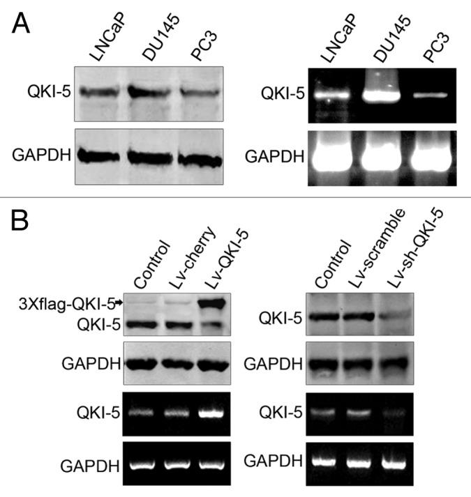
Figure 3. QKI-5 expression in PCa cell lines before and after infection with lentivirus Lv-QKI-5 and Lv-sh-QKI-5. (A) LNCaP, DU145, and PC3 cells were collected for the extraction of protein and mRNA and analyzed for QKI-5 expression using western blot and RT-PCR. Western blot and RT-PCR results showed that among the three prostate cancer cell lines, the PC3 cells had the lowest and DU145 had the highest expression of QKI-5. (B) The QKI-5 low-expressing PC3 prostatic cancer cells were infected with a lentivirus carrying QKI-5 (Lv-QKI-5) or the negative control, (Lv-cherry), and the QKI-5 high-expressing DU145 prostatic cancer cells were infected with a lentivirus carrying QKI-5 shRNA (Lv-sh-QKI-5) or a scrambled negative control (Lv-scramble). Thereafter, the protein and mRNA from these cells were extracted and subjected to western blot and RT-PCR analysis. GAPDH was used as a loading control.
