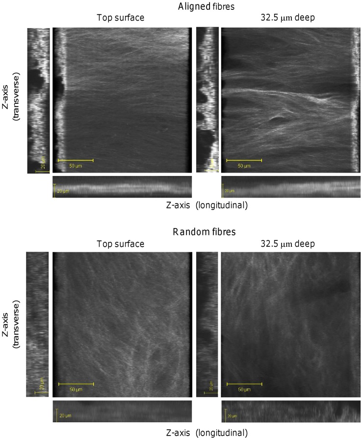
Figure 8. 3D images of collagen SHG from HDF-seeded constructs at day 21.
Images of HDF deposited-collagen on random and aligned scaffolds were compiled from 35 µm z-stacks (2.5 µm slices). A rotated 3D view from the top and bottom of the constructs are shown along with 2D images in the z-plane taken from both the horizontal and longitudinal edges.

