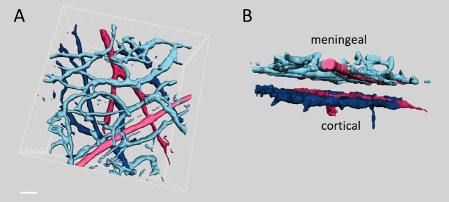
Figure 1. Upper (A) and side (B) views of 3D reconstruction of meningeal and cortical arterioles (red color) and venules (blue color) obtained from a typical z-stack of brain images.

Note the clear gap between meningeal (dural) and cortical vessels in the side view projection shown (B). Scale bar 50 µm.
