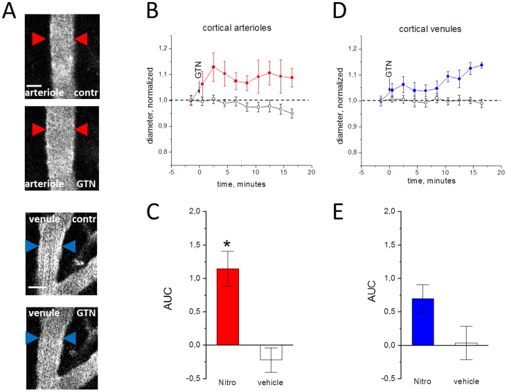
Figure 2. Effects of GTN on cortical vessels in the open cranial window preparation.
A, examples of cortical vessels before (upper image) and after GTN administration (lower image). B, the normalized diameter of cortical arterioles after injection of the GTN (filled circles) or vehicle (empty circles), real diameter before application 36.1±6 µm for cortical arterioles and 38±8 µm for cortical venules. C, comparison of changes in the area under curve (AUC) in GTN versus vehicle in cortical arterioles (n = 6 and n = 3, respectively). D and E, the same for cortical venules (n = 8 and n = 4, respectively). Note that GTN significantly changed the diameter of arterioles (* = P<0.05) but not venules (P = 0.09). Scale bar 25 µm.

