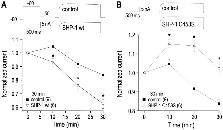
Figure 8. The hEAG1/KCNH1 current is regulated by SHP-1 tyrosine phosphatase.
The voltage protocols and normalization procedures were the same as in Fig. 4 (only traces at +60 mV are shown). A. Time-dependent effects of adding 100 µg/mL recombinant wild-type SHP-1 protein to the pipette solution. Inset: representative currents from two cells, recorded 30 min after establishing the whole-cell configuration. For each cell, the peak current at +60 mV was repeatedly measured and normalized to its initial value (3–4 min after establishing a recording) for the number of cells indicated (*p<0.05). B. Comparison of cells recorded with control pipette solution with those containing a 3∶1 mixture of the inactive substrate-trapping SHP-1 mutant (SHP-1 C453S; 150 µg/mL) and active wild-type SHP-1 (50 µg/mL). Inset: representative currents recorded 30 min after establishing the whole-cell configuration. The normalized peak currents at +60 mV are shown; *p<0.05.

