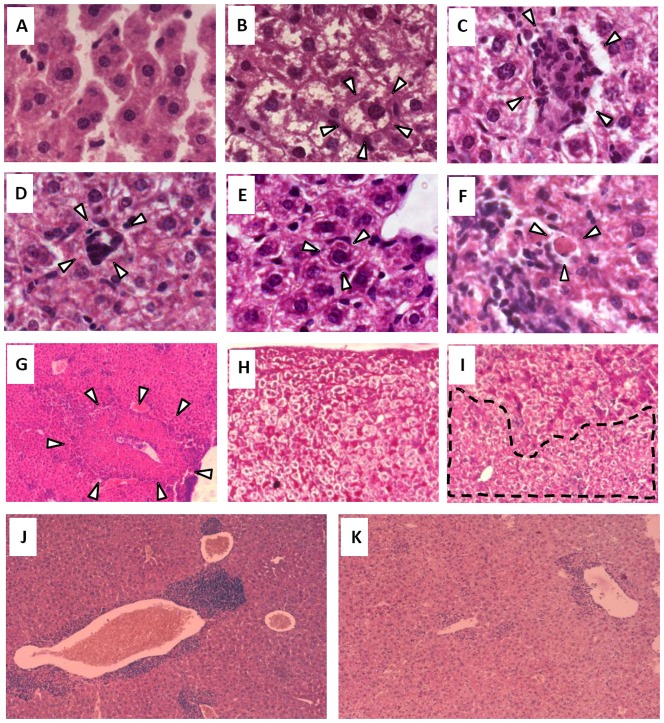
Figure 5. Histopathological alterations in hepatic tissue after systemic expression of IL-12 or IL-12+IL-18.
Livers obtained from mice in the different groups were obtained on day 14-16 post-cDNA treatment. Sections of 6 µm were obtained and (A–G) hematoxilin/eosin (H/E) or (H–I) Periodic acid-Schiff (PAS) stains were applied to the sections. (A) Hepatocytes and sinusoids from a control mouse, 100×. (B) Ballooning hepatocytes and absence of hepatic sinusoids in an IL-12-cDNA treated mouse, 100×. (C) Focus of ectopic hematopoiesis in an IL-12-cDNA treated mouse, 100×. (D) Megakaryocyte in an IL-12-cDNA treated mouse, 100×. (E) Mallory and (F) Councilman bodies in an IL-12-cDNA treated mouse, 100×. (G) Focus of necrosis; 10×. Glycogen deposits obtained in a mouse from (H) the control group or (I) the IL-12-cDNA group, 20×. Liver sections from (J) WT or (K) TNFR1 KO mice 15 days after IL-12 cDNA treatment. Pictures are representative of 2 different experiments with 4–5 mice per group.

