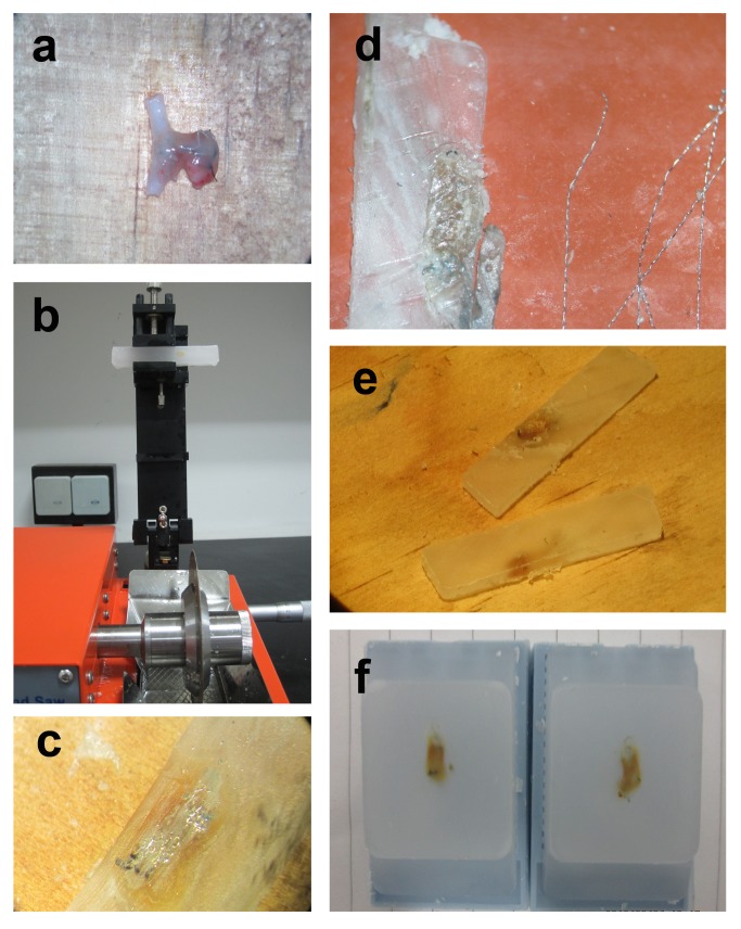
Figure 1. Embolization aneurysm specimen handling process.
(a) specimens obtained after cell transplantation for about 6 weeks (b) tissues were sectioned using an isomet low speed saw at 1000 µm intervals in a coronal orientation after being dehydrated and embedded (c), the tissue slices contained a large amount of coils under the microscope (d), the coils were extracted carefully by micro-tweezers under the microscope (e), after coil extraction and re-embedding, the tissue sections were processed into 5 mm slices by a microtome.

