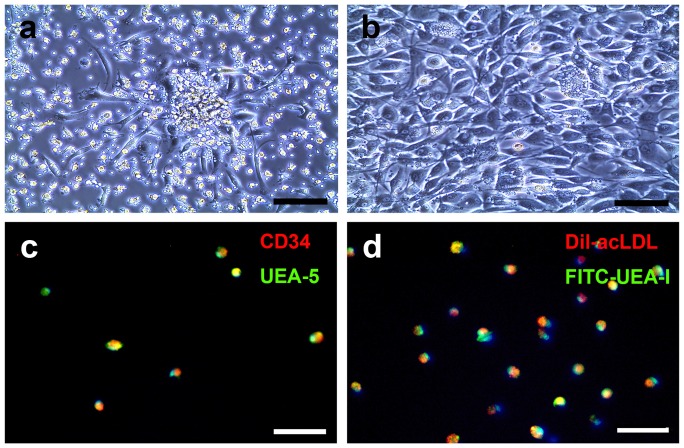
Figure 2. BM-EPC culture and identification.
A. Photomicrographs showing that after 5 days of culturing, BM-EPC isolated from rat bone marrow, displayed spindle-like attached cells and formed colonies (a), Bar = 140 µm. Higher magnification showed that these cells performed endothelial cell morphology (b), Bar = 140 µm. B. Putative BM-EPCs were stem cell marker CD34+ (red color) and were able to take in Ulex europeus agglutinin-5 (green color. c). They also were DiI-acLDL+(red color) and fluorescein isothiocyanate-Ulex europeus agglutinin-1+(green color), which were (d) co-localized in >95% cells, Bar = 100 µm.

